If you speak French, you can listen to “La Méthode Scientifique”, a program on the France Culture radio which highlighted our recent article about the ultrastructure of the periodic axonal actin scaffold. Here’s the clip with an interview of Christophe:
Our work on the ultrastructure of the periodic actin/spectrin scaffold along axons is out in Nature Communications. It’s a collaboration with platinum-replica electro microscopy specialist Stephane Vassilopoulos from the Myologie Institute in Paris.
It’s easier to go there to read it (and it’s open access!)
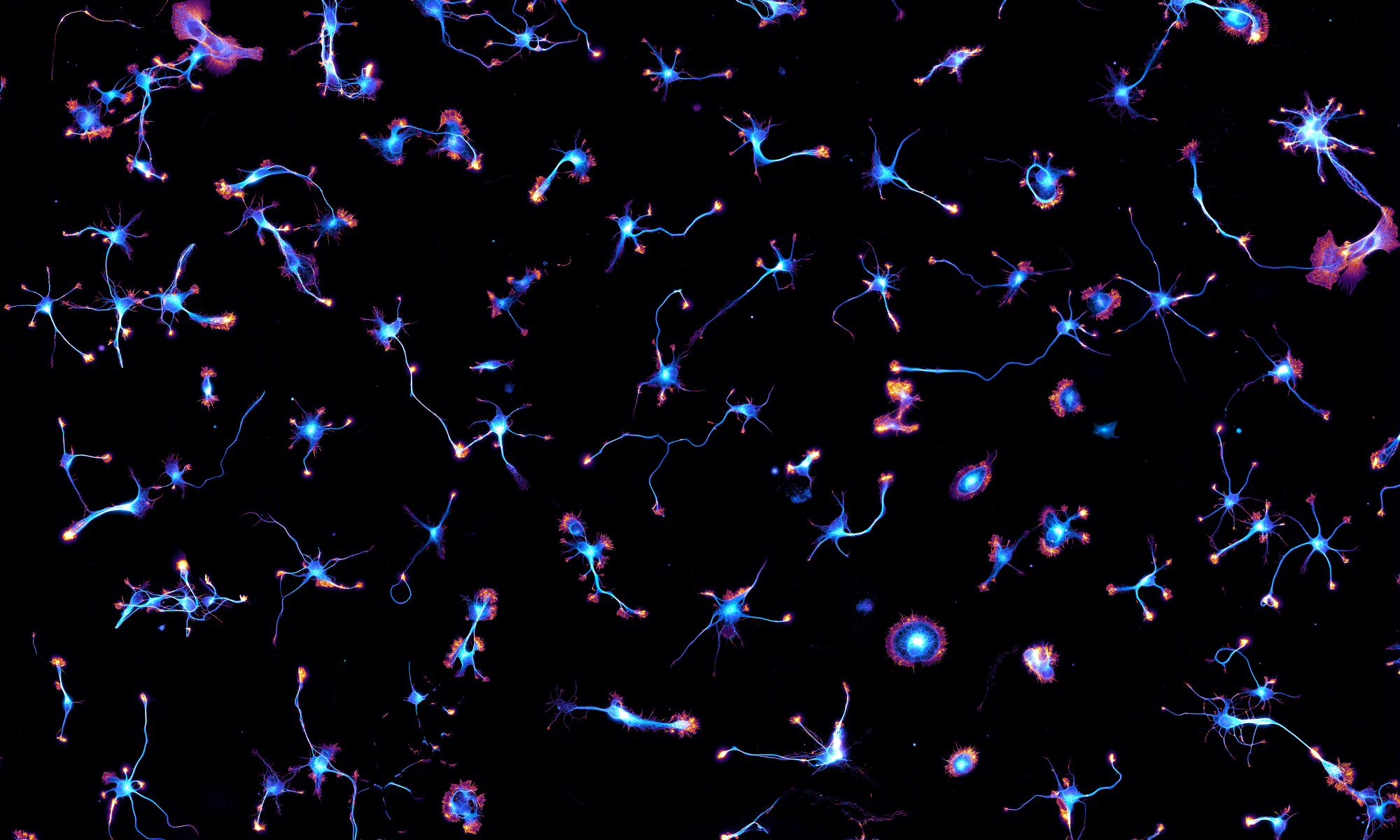
In this work that was made available as a preprintback in May, we used ultrasonic unroofing to expose the submembrane cytoskeleton along axons in neuronal cultures. This allowed to observe it both by optical super-resolution microscopy and by platinum-replica electron microscopy, zooming down to individual proteins and actin filaments.
We could visualize for the first time by EM the periodic submembrane scaffold along axons, formed of actin rings connected by spectrin tetramers. Moreover, we discovered that actin rings are not made of small actin filaments bundled together as previously assumed, but by braids of long filaments that are likely to result in their stability and flexibility. Finally, we directly visualized elements of the periodic scaffold (actin, spectrins, myosin, ankyrin) using correlative super-resolution microscopy and platinum-replica electron microscopy.
Zooming from a cultured neuron to single ankyrins along the axon initial segment by correlative STORM/PREM
A press release from CNRS is available here in English and here in French for more details about this work. We are very happy to see it out!
For the last two weeks, we got to play with Nikon Instruments latest super-resolution spinning disk microscope that incorporates a Yokogawa SoRa head. It was installed in the INP NeuroCellular Imaging Service (NCIS) imaging facility.
The Nikon SoRa setup
We could make 3D-stacks and live-cell imaging movies of cells and neurons benefiting form the ~120 nm lateral resolution. See the how this compares to diffraction-limited imaging with this example from our samples:
Comparison between raw spinning disk, raw SoRa, and deconvolved SoRa images of a COS cell labeled for actin, microtubules, clathrin and DNA.
Now that the Institute of Neurophysiopathology is settled as a whole on the Timone Campus of the AMU School of Medicine, we organized the first INP day at the Black Rock pub in Marseille. It was a big success with lots of fun and games! There were more scientific parts such as the 3-minute presentation by a PhD student of each team – Dominic did great for NeuroCyto.
The team leaders also recorded short “elevator pitches” that will be used to advertise to work of the INP teams – here’s a short behind-the-scenes video with Christophe explaining what we do:
This Friday, our new PhD students Karoline Friedl and Florian Wernert presented their projects to the members of our Institute of Neurophysiopathology (INP). They did well, and we can’t wait to see what they will do for the next three years!
Karoline presents the DAISY super-resolution techniqueFlorian present his PhD project
Christophe was invited to Cambridge by Kristian Franze and gave a seminar as part of the Adrian Seminar Series of the Department of Physiology, Development and Neuroscience. Thanks Kristian for the invitation and the glimpse to the splendid Cantabrigian life!
Saint John’s College in the morning
The next day, Christophe gave a seminar at the Chemical Engineering department, invited by Clemens Kaminski from the Laser Analytics Group. This was followed by a lively discussion with the group’s PhD students.
Christophe was invited by Benoît Charlot to speak at the scientific days of the Montpellier LabEx NUMEV, which is devoted to fostering interaction between computational and life sciences. It was an interesting experience to share our work with theoretical and computational scientist who had sometimes surprizing questions and often a fresh point of view!
On November 4th, a one-day symposium was organized at the College de France in Paris to celebrate the 10 years of the ATIP-AVENIR program, which funded and helped the creation of the NeuroCyto lab in 2017. We could hear about the history and evolution of the program from its creators and directors, as well as science stories from past awardees. Check more photos of the event here.
Christophe tries to hang a poster summarizing what the lab is about…Success for the images-as-business cards part of the poster!
Organized by the French Society for Cell Biology (SBCF), the biennal international conference “Building the Cell” is now called “Cell la Vie!”. The 2020 edition will happen from September 23rd to 36th at Institut Pasteur in Paris. Check the beautiful poster that was made from one of our images (COS cells labeled for microtubules and actin)!
Christophe spent 10 days in Bordeaux as an instructor for the FENS CAJAL Advanced Neuroscience Training Programme Course called “Advanced Techniques for Synapse Biology“. Every morning were talks from keynote speakers and instructors, while the afternoon were devoted to working on a project with two students. We heavily used the microscopes from the Bordeaux Imaging Center (thanks Magali!) including spinning disk for live-cell imaging and STORM super-resolution microscopy. The hard work of students Pushpa Khanal and Alexandra Reichova led to beautiful images of axonal trafficking and structure!
The STORM micrsocope in actionAn image from the final presentation of Pushpa and AlexandraThe course students and instructors