After doing his Master 2 with us, Florian Wernert got a PhD fellowship from École Doctorale and just started as our newest PhD student. In the next three years, Florian will use his skills in neuroscience and data analysis to dissect the organization of axonal actin structures. Welcome to the NeuroCyto team!
Together with Tijana Jovanovic-Talisman (City of Hope) and Paul Maddox (UNC), Christophe was invited by Aki Kusumi and Taka-aki Tsunoyama at the Okinawa Institute of Technology (OIST) for a seminar on new microscopy techniques. Aki and Taka were wonderful hosts and the OIST is a stunning place to do science!
A 180° view of the OIST entry gateGroup pic in front of the Kusumi lab (beautiful platinum-replica EM from Morone et al. JCB 2006)Christophe’s talk at OIST – pic courtesy of @paul_s_maddox
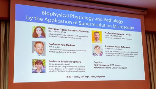
Taka and Aki then brought us on the island of Kyushu, the southern island of the main Japanese archipelago. We went to the city of Miyazaki to participate to the 57th Annual Meeting of the Biophysical Society of Japan. Our symposium was entitled “Biophysical Physiology and Pathology by the Application of Superresolution Microscopy” – featuring interesting talks with beautiful images from Takahiro Fujiwara (Kyoto University) and Makio Tokunaga (Tokyo Insitute of Technology).
We have a new PhD student! Karoline Friedl has been in the lab since March, through our partnership with the startup Abbelight.
She just now started her PhD project as a CIFRE PhD in the NeuroCyto team, in collaboration with Abbelight. She will work on Abbelight’s SAFe 360 module to develop innovative and robust multicolor nansocale imaging approaches – can’t wait to see what she comes up with!
Christophe just published a commentary about the latest article from Damaris Lorenzo and Van Bennet’s labs in PNAS. This work shows how ß2-spectrin is not only a crucial component of the periodic actin/spectrin scaffold along axons, but also directly participates in axonal transport by associating with intra-axonal vesicles.
Check it out in more details here: Leterrier C, A dual role for βII-spectrin in axons. Proceedings of the National Academy of Sciences, 2019 Jul 30;116(31):15324-15326. doi: 10.1073/pnas.1909789116
Christophe gave a course as part of the 2019 edition of the Utrecht Summer School on Neuronal Circuits Development and Plasticity. In addition to the course, there was an opportunity to discuss with the students in small groups about our science but also careers and how open science is changing the landscape – the future scientists are full of ideas and motivation!
Two visits to our neighbors at the begining of July: on July 1st, Christophe was invited by Jean-Bernard Manent to give a talk at the Institut de Neurobiologie de la Méditerranée (INMED) in Luminy. An exciting day in the artful institute, exchanging ideas with great scientists and colleagues!
On July 11th, Christophe was part of the “Advanced Photonic Imaging in Neuroscience” (APIN) symposium organized by our next-door neighbors at the Institut des Neurosciences de la Timone (INT). The symposium was filled with incredible talks showing how deeper, faster and more functional imaging are crucial to today’s neuroscience. Congrats to Ivo Vanzetta, Franck Debarbieux and Nicolas Wanaverbecq for organizing such a great event.
The Neurophysiopathology Institute, the INP we’re part of, has moved to its new headquarters on the Timone Campus of the Medicine School. It was a lot of work but everything went well and we’re almost ready to science again! Our renovated floor in the historic Medicine School building now hosts the lab and the Neuro-Cellular Imaging Service (NCIS), the new imaging facility of INP.
Christophe presented our results at the #MNS2019Mediterranean Neuroscience Society meeting in Marrakech in the “Molecular Mechanisms of Neuronal Trafficking” symposium. Thanks to Nicolas Vitale and Maïté Montero for the invitation and for organizing this!
Christophe was at the 10-year anniversary retreat of the Cytomorpho lab right in front of the sea in Les Goudes, Marseille. Thanks for the invitation Manuel and Laurent, it was great discussing with actin and microtubules specialists, and hearing the various paths taken by the lab alumni.
Marseille hosted the 2019 French neuroscience meeting from May 22 to May 24. The INP institute was present with numerous talks and posters showcasing the work of our labs (see here for a complete list). The NeuroCyto team was of course there, with Dominic presenting his first poster on the role of presynaptic actin, as well as a talk from Christophe on the ultrastructure of axonal actin rings that he also presented the day before at the satellite meeting on the spinal chord .