

We’re over the moon to see our latest work with Stéphane Vassilopoulos and his team published in Science. Check out how we reveal the unique architecture of clathrin-coated pits and endocytosis at the axon initial segment!
Don’t have access? Just email me or use this invited link to access the pdf without a subscription.

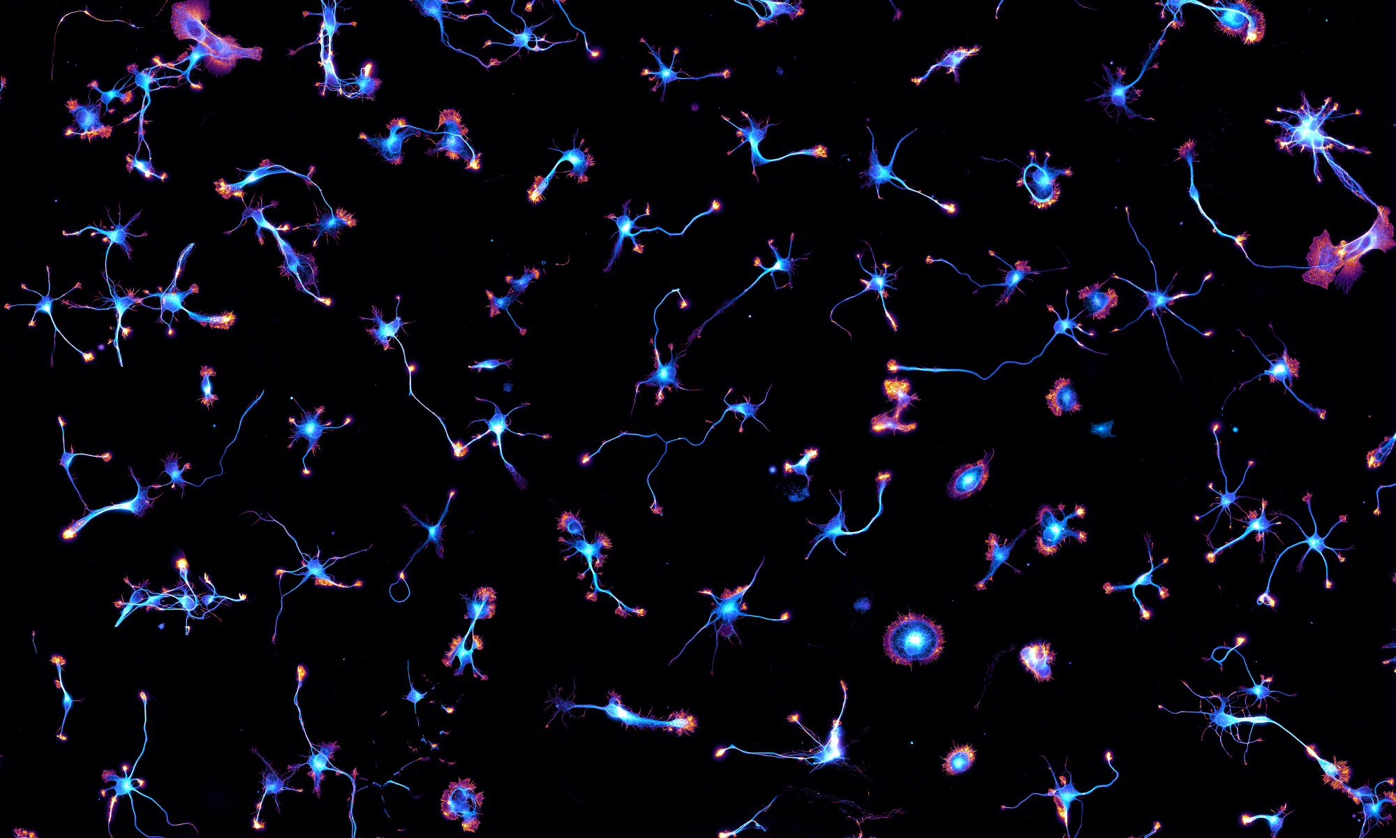
What did we discover? When we started using super-resolution microscopy and platinum-replica electron microscopy (PREM) to reveal the ultrastructure of the periodic actin-spectrin scaffold along proximal axons, we were surprised to see many clathrin-coated pits along the plasma membrane of the AIS on PREM images. Clathrin-coated pits form at the center of unique circular area of the periodic actin-spectrin scaffold that expose the bare plasma membrane: we named these exclusion area “clearings”, like in a forest.

We characterize these clearings using Structured Illumination Microscopy (SIM), Single Molecule Localization Microscopy (SMLM), PREM, and their correlative combination. Messing with axonal spectrins (using RNA interference or a drug called diamide) disrupts the periodic scaffold and results in more pits along the AIS, demonstrating that the spectrin mesh regulates pit formation. This is in line with previous findings that the spectrin scaffold can negatively define the localization of endocytic activity in epithelial cells and fibroblasts, as well as negatively regulate the endocytosis of cannabinoid receptors along axons.

When we tried to monitor endocytosis from these pits, there was another surprise! Dextran feeding resulted in most dextran cluster being present at the surface in clathrin-coated pits rather than inside the axon. This suggests that the pits are super stable and indeed, two-color TIRF-SIM of spectrin and clathrin along the AIS showed clathrin-coated pits staying for tens of minutes inside clearings:
So, why would these clathrin-coated pits form and just stay there? We show that they provide “on-demand” endocytosis: long-term depression-like stimulation with NMDA results in the scission of pits via the polymerization of “actin nests” within clearings, triggering endocytosis. This novel, regulated endocytosis mechanism makes a lot of sense at the AIS, as it allows to trigger rapid endocytosis within the otherwise super-stable submembrane scaffold. This might be how sodium channels are endocytosed from the AIS in plasticity situations to adjust neuronal excitability.

This was a blast to work on this with Stéphane, PhD student Florian Wernert and the rest of the NeuroCyto team (Florence Pelletier, Eline Simons, Fanny Boroni-Rueda, Nicolas Jullien, Marie-Jeanne Papandréou), post-doc Satish Moparthi, Jeanne Lainé, Gilles Moulay and Sofia Benkhelifa-Ziyyat in Stéphane’s team! It was mainly funded by the ANR “ASHA” we have together with Stéphane, as well as equipments grants that helped set up our Nikon Center of Excellence for Neuro-NanoImaging. Looking for a summary of our findings in French? Check the CNRS Biologie website!


