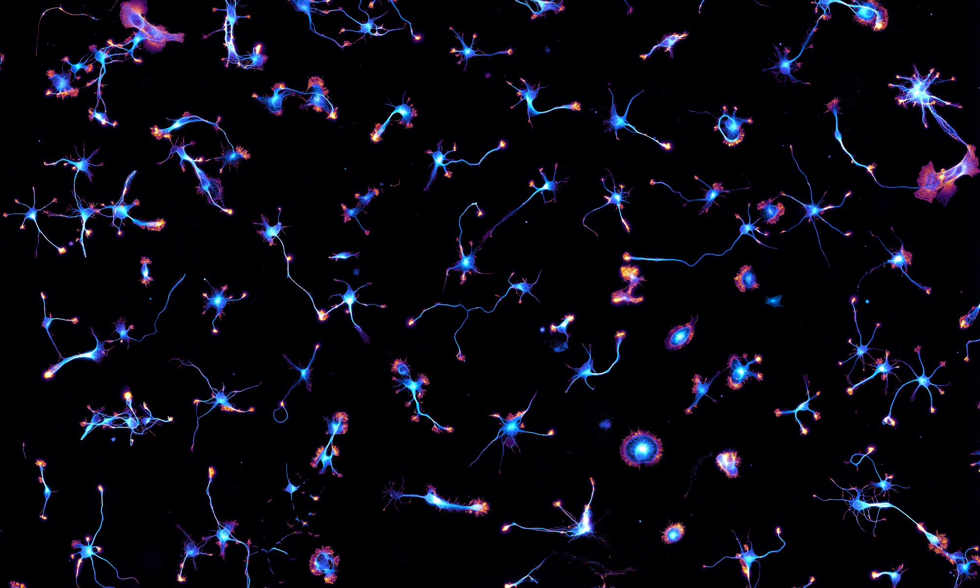
An exciting project from the lab is now out on bioRxiv. This is a collaboration with electron microscopy guru Stéphane Vassilopoulos from the Myologie Institute in Paris. One of the most striking discovery of super-resolution microscopy so far has been the periodic actin/spectrin scaffold along axons, with actin rings spaced every 190 nm by spectrin tetramers. Since its discovery in 2013, a number of labs, including ours, have used various super-resolution microscopy techniques (SMLM, SIM, STED) to refine our knowledge about this structure. However, actin rings and the axonal periodic scaffold had not been observed by EM until now.

Using mechanical unroofing and platinum-replica electron microscopy (PREM), we were able to visualize the axonal actin rings regularly spaced under the exposed plasma membrane of axons, and to determine their ultrastructure and molecular organization. And we had a big surprise: the actin rings are not made of short, capped actin filaments as it was assumed since their discovery, by analogy with actin inside the erythrocyte submembrane cytoskeleton. Instead, they are braids made of two long (~0.5 to 1 µm) actin filaments!


These actin braids are connected by a dense mesh of spectrins, which we unambiguously identified using immunogold labeling and PREM. Moreover, we probed the stability of the axonal periodic scaffold using actin-targeting drugs, and found similar results by super-resolution and electron microscopy. Finally, we directly demonstrated the identity and organization of the scaffold components (actin, spectrins) by performing correlative SMLM/PREM, visualizing the same sample by super-resolution and electron microscopy.

It was a great pleasure to work with Stéphane! The project benefited from the magic unroofing touch of our Master 2 student Solène, and from important ground work by Angélique and Ghislaine from the team. To read more, please see our bioRxiv preprint and let us know what you think!


One Reply to “New preprint: the ultrastructure of the axonal actin rings revealed!”
Comments are closed.