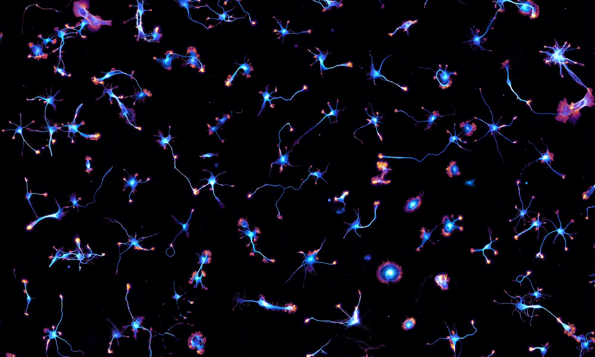
Christophe was invited to the Weizmann Institute (Rehovot, Israel) for a Molecular Neuroscience Forum seminar. Beautiful Institute, lots of interesting discussions with neuroscientists and microscopists all around the Weizmann. Thanks Nicolas Panayotis and Mike Feinzilber for the invitation!
Talks in Paris
Christophe gave two talks in Paris at the end of November, one at the Institut de la Vision in Paris, invited by Romain Brette for a PhD committee. Romain is studying how AIS position is regulated by neuronal morphology to optimize the electrogenic properties of each neuron, and he is interested by a lot of other things about the brain and its secrets as you can read on his blog.
The other talk was at a meeting at the meeting of the “Team Samples” from the RT-MFM CNRS technology group. There were interesting discussions on how to properly prepare samples for STORM and DNA-PAINT with the French super-resolution microscopy community.
Journal of Neuroscience cover
The two back-to-back papers with the Rasband lab (see more details here) have been published in the Journal of Neuroscience November 22th issue and we got the cover!
Ricardo visits us in Marseille
Ricardo Henriques, our collaborator for super-resolution stuff, including new software (see here), was in Marseille for a visit. He gave a great seminar and we got to play with Pumpy McPumpface, the Lego/Arduino-based fluidics system his PhD student Pedro Almada built.
Christophe In Amsterdam and Utrecht
Christophe was invited to the Royal Netherlands Academy of Arts and Sciences, for a colloquium on “Cell Biology of the Axon: Progress Made and Promises Ahead“. Two days discussing broads topics including axon development, axonal organization, neurodegeneration… A short Twitter thread can be found here. He was then in Utrecht invited by Amélie Fréal in Casper Hoogenraad’s lab (see the yeti-type photographic evidence from Mithila Burute below 😉). Thanks for the invite guys!
Just out: The nano-architecture of the axonal cytoskeleton
Our review on the nano-architecture of the axonal cytoskeleton is out today on the Nature Reviews Neuroscience website. This was a lot of work and a lot of fun to write with Pankaj Dubey and Subhojit Roy. We tried to provide an up-to-date view that discusses recent findings such as the various axonal actin structures visualized along the axon by STORM. We also wanted to highlight the classic EM works that shaped how we think about the axonal cytoskeleton. So it’s chock-full of recent references with fancy techniques, but also beautiful classic papers. We hope it will be a pleasant reading for all!
New preprint: a mechanism for the slow axonal transport of actin
Another work in our fruitful collaboration with Subhojit Roy and his lab (now at UW Madison). In 2015 we could visualize new axonal actin structures by STORM (see our cover): stable clusters every 3-4 µm we called “hotspots” from which “trails” would rapidly assemble and disassemble along the axon. In this new preprint, trails were shown to have a slight anterograde bias (55%) and to polymerize from the surface of the hotspots, pushing the trails away. This suggested that biased dynamic trails assembly could underlie the slow anterograde transport of actin, whose mechanism is still unknown. Modelling done by Nilaj Chakrabarty and Peter Jung (Ohio University) indeed showed that the observed biased assembly and disassembly of trails would lead to a ~0.5 mm/day transport of actin, in line with earlier measurements.

Marie-Jeanne awarded a “Pépinière d’Excellence” grant
Two collaborative papers out in the Journal of Neuroscience
In collaboration with Matt Rasband’s lab in Houston, we characterized the α-spectrin that is present along axons at the axon initial segment (AIS) and nodes of Ranvier. This work is out today as two back-to-back paper just pre-published on the Journal of Neuroscience website, here and here. Spectrins are tetramers of two α and two β subunits. It is known that the β-spectrin form at the AIS and nodes is the ßIV-spectrin since 2000, but the identity of the α subunit was unknown. In the axon, spectrins binds submembrane actin rings regularly spaced every 190 nm. As this is just below the resolution limit of conventional fluorescence microscopy (~200 nm), the resulting periodic scaffold is only visible using super-resolutive techniques such as STORM.
The first paper: “αII spectrin forms a periodic cytoskeleton at the axon initial segment and is required for nervous system function” focuses on the identification of αII-spectrin as the ßIV-spectrin partner at the AIS, and the consequences of αII-spectrin depletion in CNS-specific knockout mice. We used super-resolution microscopy to show that αII-spectrin is integrated in the AIS periodic actin/spectrin scaffold that supports the axonal plasma membrane. With the αII-spectrin antibody we used, the periodicity is seen as double bands every 190 nm by STORM. When using 2-color DNA-PAINT to image αII-spectrin together with ßIV-spectrin, the doublet of αII-spectrin labeling appears on both sides of the ßIV-spectrin bands, resolving the organization of the spectrin tetramers in situ. We also showed by STORM that the periodic actin/spectrin complex is disorganized in αII-spectrin-depleted neurons.

The second paper: “An αII spectrin based cytoskeleton protects large diameter Myelinated axons from degeneration” focuses on αII-spectrin in the PNS and nodes of Ranvier. In C. elegans mutants, the submembrane spectrin scaffold is necessary for the mechanical resistance of axons. Here, an αII-spectrin knockout mouse specific to peripheral sensory neurons was used to demonstrate this for in a vertebrate. Using STORM, we showed that loss of αII-spectrin causes a disorganization of the periodic scaffold at and around nodes. This disorganization ultimately results in the degeneration of large-diameter peripheral axons lacking αII-spectrin.

Welcome Angélique to the team
After having worked in the lab for its M1 and M2 master internships, today we’re happy to welcome Angélique Jimenez in the team as an assistant engineer. Angélique will work on various project, including bridging live-cell imaging with super-resolution microscopy to study axonal actin.


