Nikki van Bommel, a Master student from the University of Amsterdam (UvA), has joined the team for a 6-month internship, supported by an A*MIDEX fellowship. She previously worked with Joachim Goedhart (@joachimgoedhart) at UvA and wanted to learn how to do super-resolution microscopy. She will sure get plenty of training with us!
New article out: myosin II at the axon initial segment
Our collaboration with the lab of Jim Salzer (NYU) is just out. We visualized the association of phospho-myosin light chain (pMLC, an activator of the contractile myosin-II) with actin rings along the axon initial segment (AIS). This was done using two-color STORM. Moreover, acute treatment with KCL (mimicking elevated activity) resulted in a disappearance of the phospho-MLC signal before the disorganization of the actin rings. This suggests that myosin-II contractility has a role in setting the AIS shape and position along the axon, and that release of this contractility could be a key remodelling step for the activity-dependant plasticity of the AIS.

Visit to the Weizmann Institute
Christophe was invited to the Weizmann Institute (Rehovot, Israel) for a Molecular Neuroscience Forum seminar. Beautiful Institute, lots of interesting discussions with neuroscientists and microscopists all around the Weizmann. Thanks Nicolas Panayotis and Mike Feinzilber for the invitation!


Talks in Paris
Christophe gave two talks in Paris at the end of November, one at the Institut de la Vision in Paris, invited by Romain Brette for a PhD committee. Romain is studying how AIS position is regulated by neuronal morphology to optimize the electrogenic properties of each neuron, and he is interested by a lot of other things about the brain and its secrets as you can read on his blog.
The other talk was at a meeting at the meeting of the “Team Samples” from the RT-MFM CNRS technology group. There were interesting discussions on how to properly prepare samples for STORM and DNA-PAINT with the French super-resolution microscopy community.
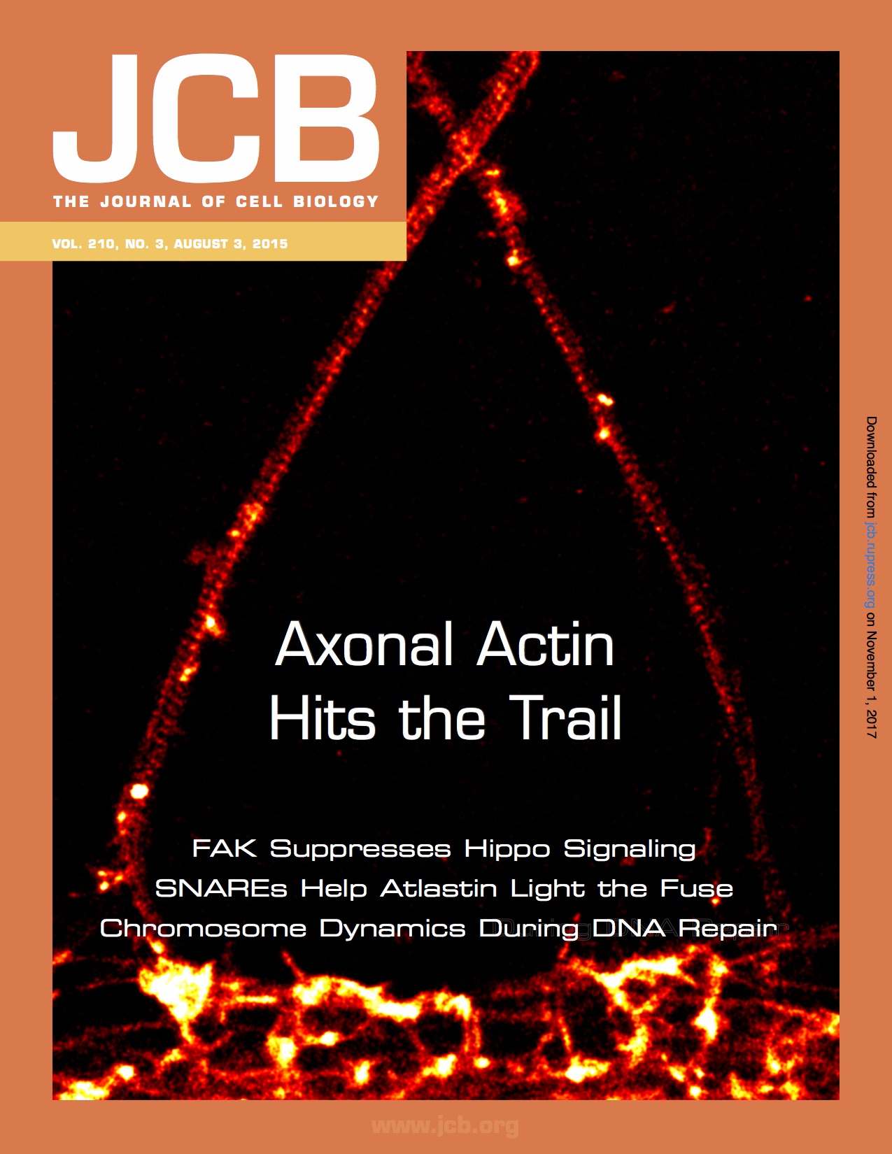
Journal of Neuroscience cover
The two back-to-back papers with the Rasband lab (see more details here) have been published in the Journal of Neuroscience November 22th issue and we got the cover!

Ricardo visits us in Marseille
Ricardo Henriques, our collaborator for super-resolution stuff, including new software (see here), was in Marseille for a visit. He gave a great seminar and we got to play with Pumpy McPumpface, the Lego/Arduino-based fluidics system his PhD student Pedro Almada built.


Christophe In Amsterdam and Utrecht
Christophe was invited to the Royal Netherlands Academy of Arts and Sciences, for a colloquium on “Cell Biology of the Axon: Progress Made and Promises Ahead“. Two days discussing broads topics including axon development, axonal organization, neurodegeneration… A short Twitter thread can be found here. He was then in Utrecht invited by Amélie Fréal in Casper Hoogenraad’s lab (see the yeti-type photographic evidence from Mithila Burute below 😉). Thanks for the invite guys!
Just out: The nano-architecture of the axonal cytoskeleton
Our review on the nano-architecture of the axonal cytoskeleton is out today on the Nature Reviews Neuroscience website. This was a lot of work and a lot of fun to write with Pankaj Dubey and Subhojit Roy. We tried to provide an up-to-date view that discusses recent findings such as the various axonal actin structures visualized along the axon by STORM. We also wanted to highlight the classic EM works that shaped how we think about the axonal cytoskeleton. So it’s chock-full of recent references with fancy techniques, but also beautiful classic papers. We hope it will be a pleasant reading for all!

New preprint: a mechanism for the slow axonal transport of actin
Another work in our fruitful collaboration with Subhojit Roy and his lab (now at UW Madison). In 2015 we could visualize new axonal actin structures by STORM (see our cover): stable clusters every 3-4 µm we called “hotspots” from which “trails” would rapidly assemble and disassemble along the axon. In this new preprint, trails were shown to have a slight anterograde bias (55%) and to polymerize from the surface of the hotspots, pushing the trails away. This suggested that biased dynamic trails assembly could underlie the slow anterograde transport of actin, whose mechanism is still unknown. Modelling done by Nilaj Chakrabarty and Peter Jung (Ohio University) indeed showed that the observed biased assembly and disassembly of trails would lead to a ~0.5 mm/day transport of actin, in line with earlier measurements.
