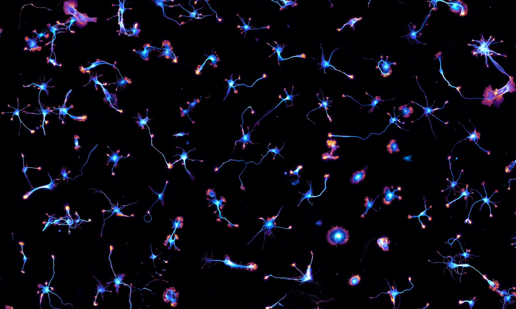
Our work on the ultrastructure of the periodic actin/spectrin scaffold along axons is out in Nature Communications. It’s a collaboration with platinum-replica electro microscopy specialist Stephane Vassilopoulos from the Myologie Institute in Paris.

In this work that was made available as a preprint back in May, we used ultrasonic unroofing to expose the submembrane cytoskeleton along axons in neuronal cultures. This allowed to observe it both by optical super-resolution microscopy and by platinum-replica electron microscopy, zooming down to individual proteins and actin filaments.
We could visualize for the first time by EM the periodic submembrane scaffold along axons, formed of actin rings connected by spectrin tetramers. Moreover, we discovered that actin rings are not made of small actin filaments bundled together as previously assumed, but by braids of long filaments that are likely to result in their stability and flexibility. Finally, we directly visualized elements of the periodic scaffold (actin, spectrins, myosin, ankyrin) using correlative super-resolution microscopy and platinum-replica electron microscopy.

A press release from CNRS is available here in English and here in French for more details about this work. We are very happy to see it out!


One Reply to “Our paper is out! The ultrastructure of the axonal actin rings revealed”
Comments are closed.