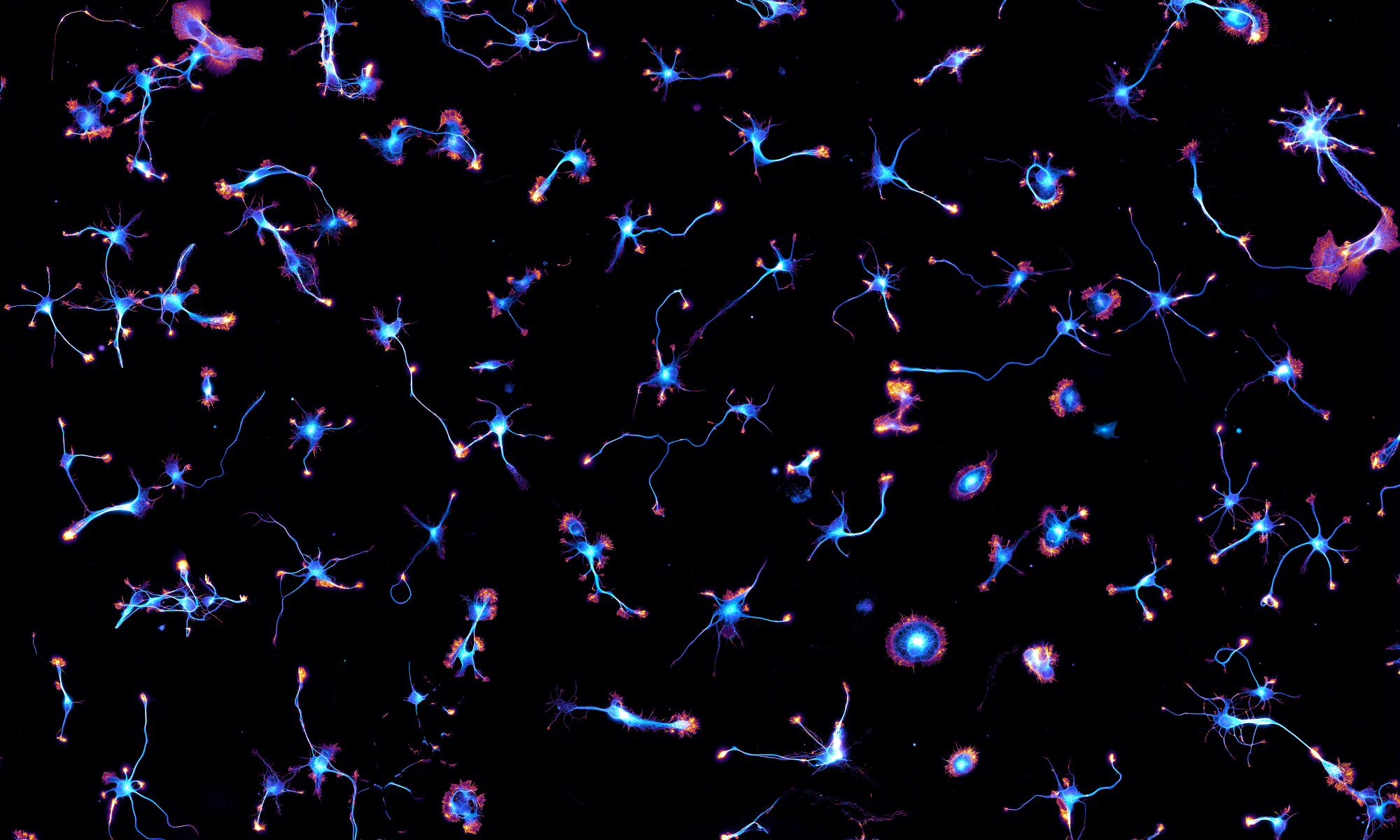
We have a new preprint out! It’s a collaboration with the group of Sandrine Lévêque-Fort at ISMO (Orsay, France) based on the PhD work of Clément Cabriel. They previously used supercritical-angle fluorescence to measure the height of fluorophores at the proximity of the coverslip. This technique, called Direct Optical Nanoscopy with Axially Localized Detection (DONALD), could bring the resolution down to 15 nm for 3D localization microscopy. Now they have combined the SAF-based method with cylindrical lens astigmatism to obtain a robust and precise 3D localization of fluorophores over ~1.5 µm above the coverslip, retaining the key advantafges of DONALD: drift-free, tilt-insensitive and achromatic. The new technique, called Dual-view Astigmatic Imaging with SAF Yield (DAISY 😉), allowed to image in 3D the periodic scaffold of adducin and ß-spectrin along axons of cultured neurons, as you can see on the Figure below:


