We had the chance of having Dominic Bingham, currently a Master’s student in Alison Twelvetree’s lab in Sheffield, apply for a PhD in the lab through the “Integrative and Clinical Neuroscience” PhD program. Congratulation to Dominic who succeeded to be one of the three laureates this year! We can’t wait to have him the lab where he will study the organization of presynaptic actin. Thanks also to the Aix-Marseille University A*MIDEX which is funding the PhD program.
Christophe talks at the French Cell Adhesion Club meeting in Strasbourg
It was a pleasure to present at the 7th Cell Adhesion Club Meeting in Strasbourg. After three fascinating days hearing about cell adhesion and related cell biological processes by top scientists of the field, it was very interesting to see how key tools and concepts can also inform cellular neurobiology. See the #Stradh18 Twitter hashtag for more, thanks to @GoetzJacky for the invite!


Just published: our review on the architecture of axonal actin
Marie-Jeanne and Christophe wrote a review detailing how recent discoveries renewed the understanding of axonal actin organization. In the axon shaft itself, new nano-structures such as rings, hotspots and trails have been described, but their function remains to be elucidated. At presynapses, the precise architecture of actin is still elusive, and contradicting findings have been reported regarding its function. This is an exciting time to study actin in axons!
The review is now published in Molecular and Cellular Neuroscience, and will be part of a special issue on “Membrane Trafficking and Cytoskeletal Dynamics in Neuronal Function”. If you don’t have access to the review, a preprint manuscript is available on Zenodo.

New preprint: Pumpy is here!
Together with the he lab of Ricardo Henriques, we’re finally ready to present #Pumpy to the world, in the form of a preprint on bioRxiv!

Pumpy (short for Pumpy McPumpface, official name NanoJ-Fluidics) is a pump array made in LEGO, controlled by an Arduino and open-source software (compatible with ImageJ/Fiji and Micro-Manager). NanoJ-Fluidics automates sample fluid exchange right on the microscope stage, allowing complex workflows using your standard chambers, tubing and reagents: live-to-fixed correlative imaging, sequential staining/imaging/washing protocols…
In one application, we used NanoJ-Fluidics to visualize the dynamics of actin in living cells (using SRRF super-resolved processing), and to perform nanoscale imaging of actin using STORM on the same cell after online fixation and labeling with phalloidin.

We also used NanoJ-Fluidics for sequential multiplexed STORM/PAINT imaging, and we obtained 5-channel super-resolved images of actin, intermediate filaments, microtubules, clathrin and mitochondria in cells with minimal intervention during imaging.

If you are interested in making your own, head over to the NanoJ-Fluidics wiki where you will find everything: LEGO parts, assembly instructions, control software and more!

Christophe presents at the École des Houches BIOPOL summer school
Christophe gave a conference on “The neuronal cytoskeleton revealed by cellular imaging” at the “Mechanobiology of polarized cell” summer school at the Ecole de Physique des Houches (in front of the Mont-Blanc!). This was organized by the PhD students of the Marie Curie Sklodowska International Training Network “Biochemical and mechanochemical signaling in polarized cells” (BIOPOL). A good occasion to present what super-resolution microscopy can reveal about polarized cells organization, and discuss about the mechanical implications of the axonal cytoskeleton architecture. See more from the #mechanopol18 hashtag on twitter.


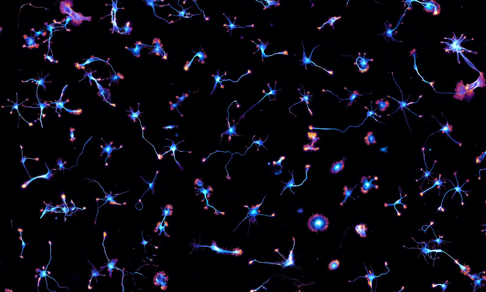
One of our neurons in the Parisian subway!


Just out: a Viewpoint on the Axon Initial Segment
Christophe wrote a short review for the Journal of Neuroscience’s Viewpoints series, summarizing the latest results about the Axon Initial Segment (AIS). It’s out today in J. Neurosience latest issue. Also, a nice image of our neurons with their initial segments was chosen as the cover!

Christophe talks at IBDM
Christophe gave a seminar at IBDM in Marseille, not too far from the lab. Thanks Francis for the invitation!
We support PreLights, a preprint highlighting initiative
Today the Company of Biologists (a non-profit organization that published journals such as Development or the Journal of Cell Science) launched a new website called PreLights. PreLights is a website where a group of young researchers (the PreLighters) curate biology preprints, highlighting their value and importance. Some highlights also feature feedback from authors. We are happy to support this open science initiative by providing the PreLight banner image: neurons in culture that reflects the connections and interactions preLights wants to promote.

The SQUIRREL is out!
 Our article with the Henriques and Mercer labs is now out in Nature Methods. Previously posted on bioRxiv, it proposes a new metric to measure the quality of super-resolution images called NanoJ-SQUIRREL (Super-resolution Quantitative Image Rating and Reporting of Error Locations). Simply put, it compares the image to a reference diffraction-limited image, allowing to detect artefacts and missing features in the super-resolved image.

We used SQUIRREL to determine when to stop a STORM acquisition, showing that the longest acquisition was not always the best. In this case of imaging actin rings along axons labeled with phalloïdin, the quality of the STORM image as measured by SQUIRREL peaks after 30,000 frames and slightly drops when reaching 60,000 frames:

Another application of SQUIRREL is to optimize a DNA-PAINT experiment. In DNA-PAINT imaging, the density of the blinking events can be tuned by varying the concentration of the imager strand. We imaged clathrin-coated pits in a glial cell and the blinking sequence were processed with different algorithms. Each algorithm had a specific optimal blinking density (and thus imager strand concentration): SRRF was better with a denser blinking sequence, while pure localization algorithms such as MLE or CoM require a sparser acquisition sequence:

There are a lot more applications in the paper and its hefty supplementary file, go check it! Then try it thanks to the easy to use, open-source plugin for the ImageJ/Fiji plugin software.
Finally, this project is a striking example of open science in action. I (Christophe) met and started to collaborate with Ricardo and Jason on Twitter; the manuscript was posted on bioRxiv, where it got feedback from beta-testers and caught the attention of editors, before being accepted for publication six months later. Congrats to Siân, David, Caron, Pedro, and everyone!