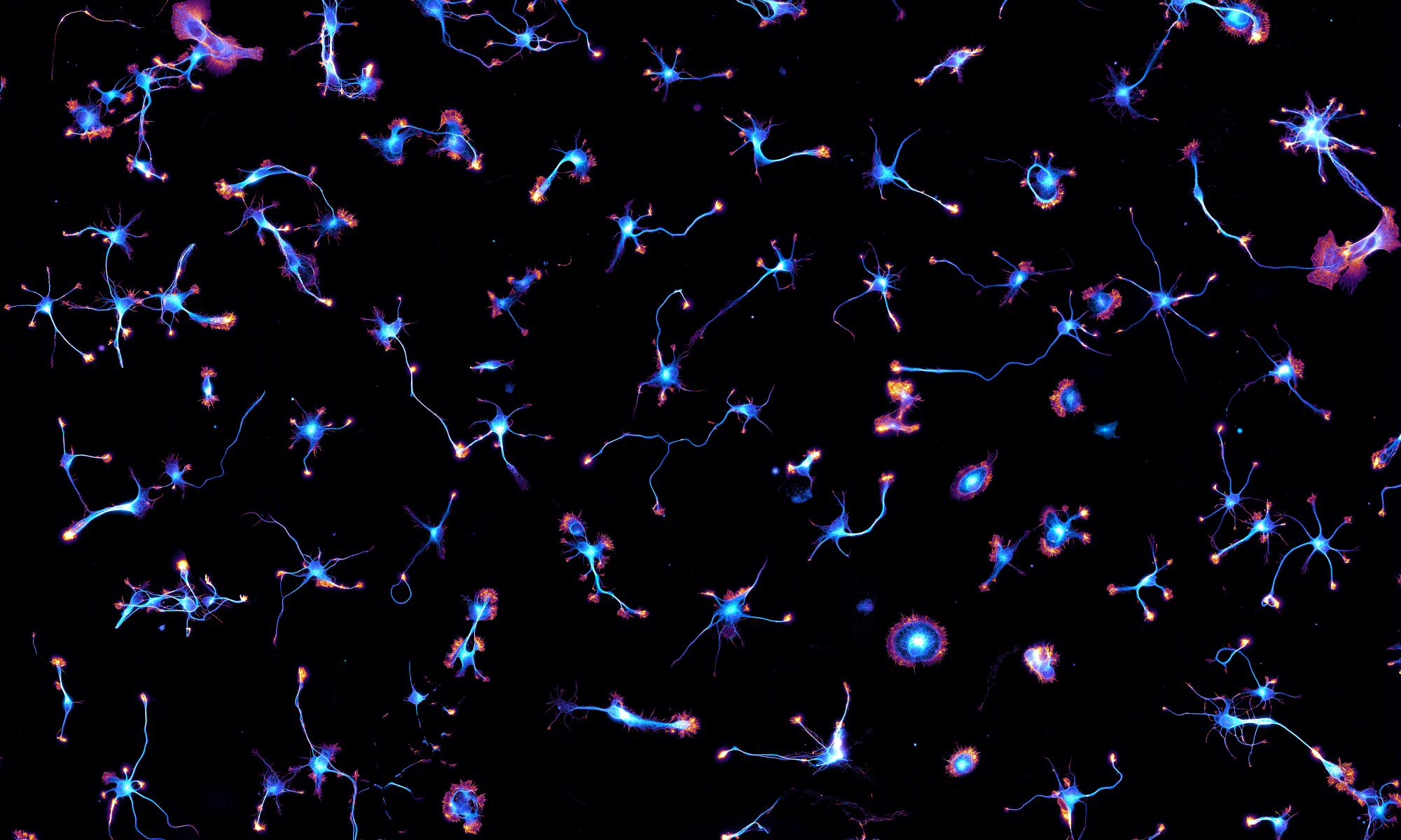
We have a
new preprint out! It’s a collaboration with the group of Sandrine Lévêque-Fort at ISMO (Orsay, France) based on the PhD work of Clément Cabriel. They previously used supercritical-angle fluorescence to measure the height of fluorophores at the proximity of the coverslip. This technique, called Direct Optical Nanoscopy with Axially Localized Detection (
DONALD), could bring the resolution down to 15 nm for 3D localization microscopy. Now they have combined the SAF-based method with cylindrical lens astigmatism to obtain a robust and precise 3D localization of fluorophores over ~1.5 µm above the coverslip, retaining the key advantafges of DONALD: drift-free, tilt-insensitive and achromatic. The new technique, called Dual-view Astigmatic Imaging with SAF Yield (
DAISY 😉), allowed to image in 3D the periodic scaffold of adducin and ß-spectrin along axons of cultured neurons, as you can see on the Figure below: